Under- & Post-Graduate Studies
At North Staffs Poly I took a Combined Sciences degree, beginning with Physics, Biology and Computing. As is the way with this course, at the start of the second year I focused on Biology and Physics.
Following the completion of my degree I took work as a laboratory technician (below) to support myself through a part-time master’s degree in comparative animal physiology at Birkbeck College, University of London.
Technical
During my MSc. studies I first worked as a laboratory technician at the Department of Physiology, Royal Free Hospital School of Medicine (now part of University College) running the laboratories of Professor K. Michael Spyer. These laboratories carried out in vivo research on the neurophysiology of respiratory control and fight-or flight mechanisms, involving recording activity in brain centres and even individual neurons using glass-filament microelectrodes .
I operated and maintained equipment such as blood-gas analysers, respiratory pumps, electronic stimulation and recording apparatus, surgical instruments and making-up salines, drugs and anaesthetics for a wide variety of procedures.
After 18 months I moved to North East Surrey College of Technology as a technician running the main physiology and pharmacology laboratories: Maintaining and building new equipment, making up solutions and drug preparations and ensuring provision of a clean, safe environment in an increasing number of laboratories and preparation areas. As staff changed I was promoted twice and covered several other teaching areas including microbiology, radiobiology & virology. With my MSc completed and the departure of the chief technician I sought research work elsewhere.
Pupillometry & Developing Vision
In 1991 I joined the research group at The University of Birmingham Academic Unit of Ophthalmology under Professor Alistair Fielder, in whose departments I would work up to his retirement in 2009 and with whom I continue to collaborate on the RetVas project (below).
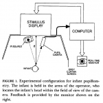
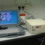
At the Birmingham and Midland Eye Hospital (now relocated to The Birmingham and Midland Eye Centre) I was tasked with assembling and operating an adult pupillometer to study the phenomenon of pupillary reactions to patterns and it’s potential use in assessing visual acuity. I established methodologies of measuring visual acuities from adults using pupillary reactions to carefully-controlled, structured stimuli.
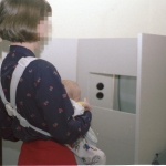
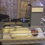
I further adapted the pupillometer for use with infants and neonates in a laboratory I established at Dudley Road Hospital Neonatal Intensive Care Unit.
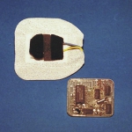
I was also involved in the development of an ‘Intelligent Eye-Patch’ for the monitoring of compliance amongst children being treated by occlusion therapy for amblyopia.
In 1995, Professor Fielder’s research group moved to Imperial College London, based initially at the Western Eye Hospital and later at Charing Cross Hospital. I continued to develop the infant pupillometer in a laboratory I established at Hammersmith Hospital NICU and succeeded in recording light reflexes and visual activities from premature infants.
I have presented my work at many national and international meetings including meetings of The Association for Research in Vision and Ophthalmology, The Pupil Colloquium and the Child Vision Research Society. I have managed the Web presence of the CVRS for the last 15 years.
Retinopathy of Prematurity (ROP)
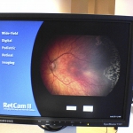
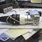
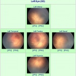
Between 1995 and 2005 I helped run the Retinopathy of Prematurity ward rounds at Imperial College’s St Mary’s Hospital Neonatal Intensive Care Unit. This involved the initial organization of visits to patients, operating the camera while Professor Fielder and other clinicians acquired pictures and maintaining upgrading and improving the equipment and methodologies used.
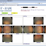
The huge numbers of images acquired during these clinics led me to realise a better way of accessing the data was needed and I developed a sever-based image database to host data from two generations of RetCam.
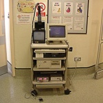
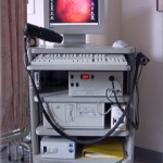
I was able to convert the trolley-mounted RetCam into a self-contained transport case that could be more easily moved to other neonatal units, including Chelsea and Westminster Hospital.
Retinal Image Analysis
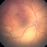
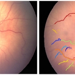
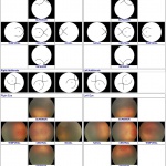
I have contributed to several projects involving the analysis of retinal images from the Imperial College RetCams between 1995 and 2005. I have written scripts and applications to compare and analyse retinal images, comparing different doctor’s assessments of imaged retinae, measuring vessel geometry and the size and separations of structures on the retina such as the optic disk, macula and ROP lesions.
Telemedicine
Telemedicine is the acquisition of medical data from a remote patient, it’s transmission to qualified personnel and the return of diagnostic conclusions for local action.
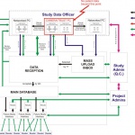
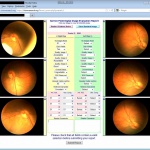
My experience in acquiring retinal images, storing them on a server and distributing them for remote assessment, led me to be invited to collaborate in the int BOOST/RIDA international study. I was responsible for writing software to anonymize data collected from RetCams in NHS neonatal units around the UK, storing the sanitized data – with the NHS Trust’s permission – on my personal server and writing web interfaces for researchers around the world to access the data and conduct studies.
My scripts gathered their responses, collated the data and presented them for analysis that formed the basis of several papers and meeting abstracts.
RetVas.com
At Imperial College London, our research group began to develop methodologies and a program to quantify parameters of vessel morphology in eyes affected to varying degrees by ROP.
A casual remark by me that I could take that program and run it on a web server was later developed by Clare Wilson into a commercial venture called RetVas.
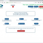
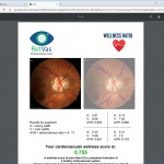
RetVas aimed to take retinal images uploaded from clinical sources and provide assessments of the vessel morphology to quantify cardiovascular health, diabetic retinopathy and ROP.
I joined the team to realize the web-based aspects of the project:-
• Establishing secure stand-alone and later cloud servers to host operations.
• A public website presenting the commercial aspect of the project.
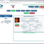
• Secure upload and storage of images, including a zero-knowledge encryption of patient identity to which only the client has access.
• Preparing and passing images to the central analysis program.
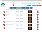
• Capture, format and present the results in a report downloadable by the originating client.
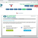
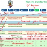
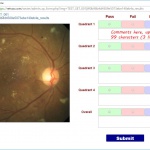
• Ensuring the system secures data, keeps backups and monitors quality and integrity of original submissions and results.
